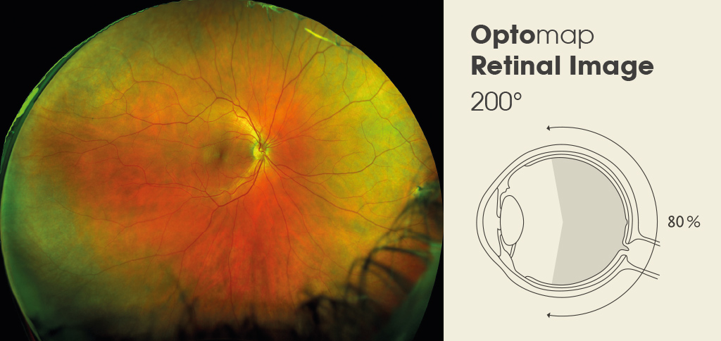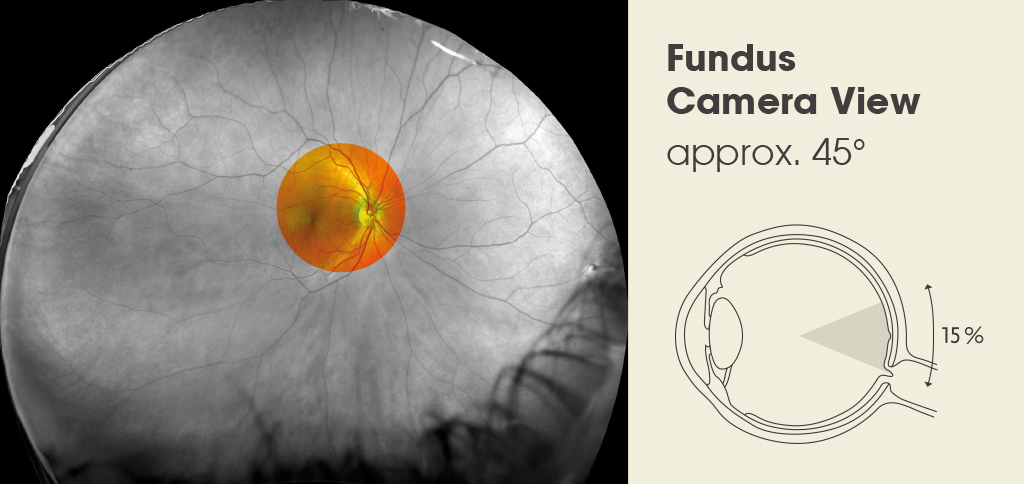
The optomap ultra-wide digital retinal imaging system captures more than 80% of your retina in one panoramic image. Traditional viewing methods typically reveal only 10- 15% of your retina at one time and are carried out manually without any digital record.
The unique optomap ultra- wide view enhances your optometrist’s ability to detect even the earliest signs of disease that appear on your retina. Seeing most of the retina at once allows your optometrist more time to review your images and educate you about your eye health. Numerous clinical studies have demonstrated the power of optomap as a diagnostic tool.
Your optomap scan includes a colour red/green image, a red free scan which images the retina (the light receptor layer), a green free scan which images the choroid (the blood rich layer behind the retina) and an auto-fluorescence (AF) scan which allows your optometrist to assess the function of the retina assisting in the diagnosis and monitoring of eye conditions.

